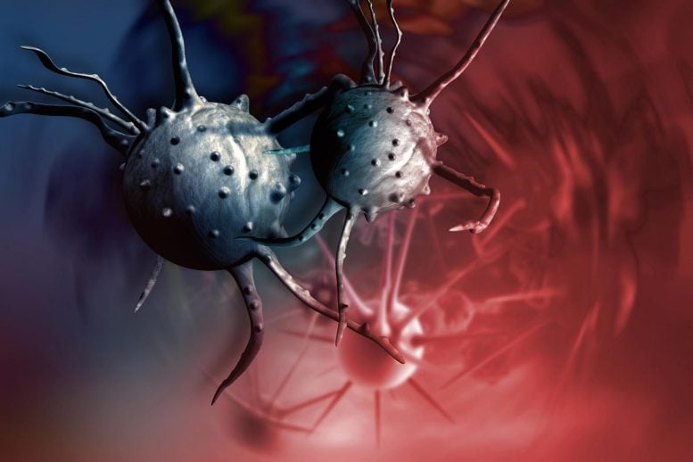
New research reveals that polyploid giant cancer cells (PGCCs), which can adapt and survive after treatment, are key to cancer recurrence. These cells change their genes to protect themselves against treatment, then divide to cause the tumor to regrow. Targeting these cells with specific inhibitors like p21 during treatment could improve cancer treatment outcomes.
Scientists have discovered that giant polyploid cancer cells, monstrously oversized and containing multiple nuclei, may be responsible for disease recurrence after cancer treatment.
Researchers at MUSC Hollings Cancer Center have made a breakthrough that could reveal why cancer sometimes comes back in patients who have received chemotherapy or radiation therapy.
Both forms of therapy aim to stress cancer cells until they self-destruct. However, these treatments often lack long-term effectiveness because cancer cells can adapt to stress, escape, and allow the tumor to rebound after a short period of time.
Recently, scientists have begun to study the role of polyploid giant cancer cells, or PGCCs, in cancer recurrence. Although these cells have been known to scientists since the invention of the microscope and have been observed by pathologists in cancerous tissues, their exact function in cancer recurrence remained unknown.
In a recent article from Journal of Biological Chemistry, a research team at MUSC Hollings Cancer Center led by Christina Voelkel-Johnson, Ph.D., reports that it has identified certain genes that prostate cancer cells manipulate to become PGCCs, thereby protecting themselves from therapeutic stress. Hollings’ team also discovered that PGCCs later regained their cell division capacity, paving the way for cancer recurrence.
Unexpected discoveries during laboratory experiments
Voelkel-Johnson and his lab made the discovery while studying an inhibitor, or a drug designed to block a biological mechanism, associated with long-lasting cures after radiation therapy. “We initially thought that combining the radiation with the inhibitor killed the cancer cells better,” Voelkel-Johnson said. “Only when the inhibitor failed to make a difference in short-term experiments was the time frame extended, allowing for an unusual observation.”
Lab members had observed abnormal-looking giant cells in short-term experiments, but considered them “doomed.” When the delay was extended, they were surprised to find that these cells generated small offspring.
This time-lapse video shows the formation of PGCC in ovarian cancer cells in response to treatment-related stress. Credit: Video courtesy of Joe. R. Delaney, Medical University of South Carolina
“They looked really awesome,” Voelkel-Johnson said. “When we weren’t using the inhibitor, these giant cancer cells generated daughter cells, creating the appearance of a colony with smaller cells surrounding the larger one.”
These funky-looking PGCCs were visually different from other cancer cells. They were able to make copies of their genetic information, thereby increasing the number of nuclei. However, the cytoplasm did not divide and the cells grew monstrously, containing several nuclei instead of just one.

Dr. Christina Voelkel-Johnson, researcher at MUSC Hollings Cancer Center. Credit: Medical University of South Carolina. Photography by Sarah Pack.
The surprise findings that the monster cells were not “doomed” led Voelkel-Johnson and his team to suspect that their inhibitor had stopped cancer recurrence in a different way than they had assumed.
“The inhibitor did not kill the cancer cells any better,” Voelkel-Johnson said. “Instead, it prevented the generation of progeny from the polyploid giant cancer cells.”
The team also observed that PGCC daughter cells continued to divide, mimicking the tumor recurrence that some patients experience after treatment. It became clear that this inhibitor created long-lasting cures not by causing cell death, but by preventing PGCCs from reverting to single-nucleus cancer cells capable of dividing.
To understand what differentiated PGCCs and their daughter cells from their parental cancer cells, Voelkel-Johnson, with the help of other collaborators, set out to study changes in gene expression among the different cells that arose during of their experiences. This information would help explain how cancer cells can enter and exit PGCC states after being exposed to therapeutic stress.
Genetic knowledge and therapeutic implications
Voelkel-Johnson and her team were able to identify the cell signaling pathways that cancer cells manipulate to become PGCCs in response to therapeutic stress, then later revert to cells capable of producing daughter cells.
One protein that particularly piqued their interest was p21, which is induced by a protein called p53 when normal cells are stressed. In normal cells, p21 prevents damaged cells from duplicating.
” data-gt-translate-attributes=”({“attribute”:”data-cmtooltip”, “format”:”html”})” tabindex=”0″ role=”link”>DNA, allowing repair of damage caused to DNA. Cells in which the damage cannot be repaired commit suicide.
Hollings’ research team showed that stress in cancer cells lacking p53 also increased p21, but the protein did not stop the duplication of damaged DNA, as it did in normal cells. As a result, p21 helped set the stage for the generation of PGCCs.
When increases in p21 were blocked, stressed cancer cells didn’t turn into these monster cells. Interfering with p21 in already monstrous cells prevented them from generating daughter cells that could be responsible for tumor relapse.
The team’s findings provide insights into novel mechanisms that could be targeted to improve patient outcomes after cancer treatment. While it may not be possible to block p21 as a treatment, the breast cancer drug tamoxifen and cholesterol-lowering statins have been shown to interfere with the pathways the team identified. Further research is needed to assess whether they can reduce recurrence rates by preventing PGCCs from regaining the ability to generate daughter cells.
The results also provide new information on the optimal timing for administration of these drugs.
“One of the questions we asked was, ‘At what point in therapy do you treat?’ “, Voelkel-Johnson said. “Our results suggest that treatment should take place at the same time as chemotherapy or radiotherapy. It is important to administer one of these drugs in combination with therapeutic stress to prevent PGCCs from generating the daughter cell. Once they are generated, it is too late.
Voelkel-Johnson plans to continue investigating ways to prevent the generation of daughter cells from PGCCs to increase the effectiveness of the treatment. She is also interested in evaluating how various combination regimens given at the time of cancer treatment affect recurrence rates in a wide range of cancers.
Reference: “Transcriptome analysis of polyploid giant cancer cells and their progeny reveals a functional role for p21 in polyploidization and depolyploidization” by Shai White-Gilbertson, Ping Lu, Ozge Saatci, Ozgur Sahin, Joe R. Delaney, Besim Ogretmen and Christina Voelkel-Johnson, March 4, 2024, Journal of Biological Chemistry.
DOI: 10.1016/j.jbc.2024.107136
The study was funded by the National Cancer Institute, the
” data-gt-translate-attributes=”({“attribute”:”data-cmtooltip”, “format”:”html”})” tabindex=”0″ role=”link”>National Institutes of Healthand the American Cancer Society.