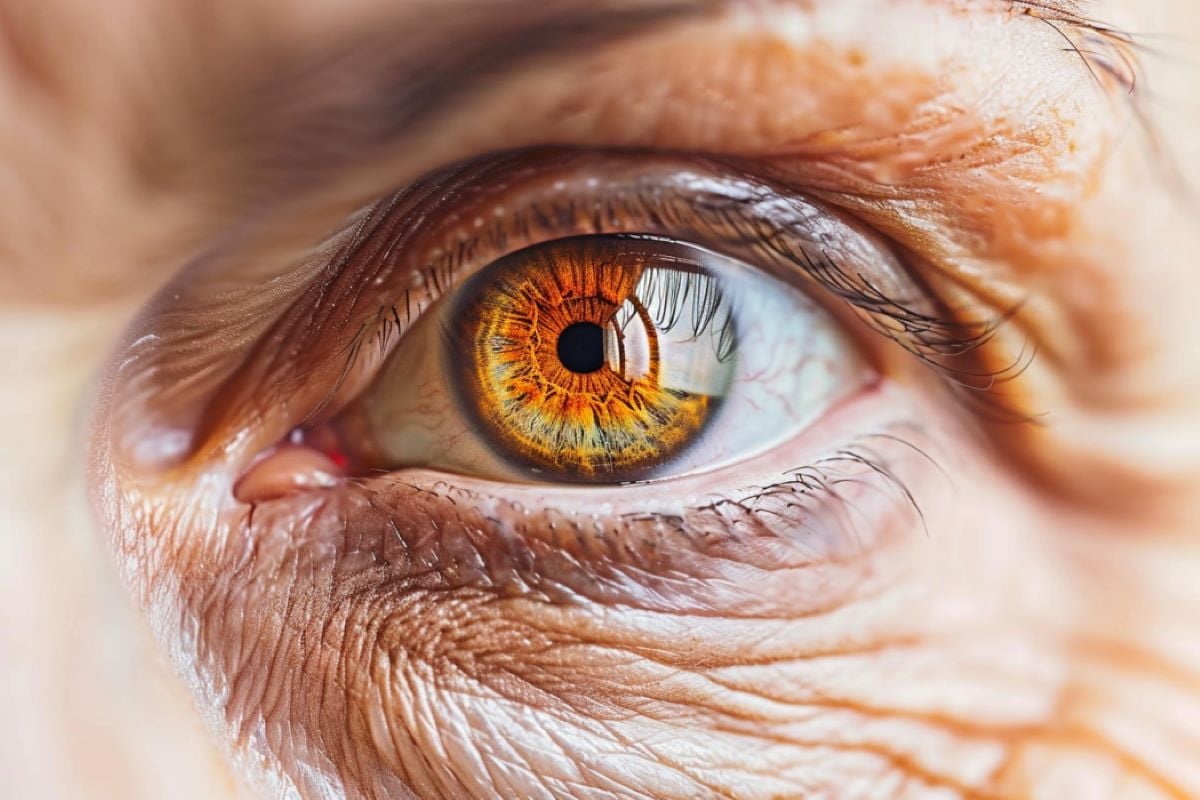
Summary: Researchers have found that daily antioxidant supplements slow the progression of late-stage dry age-related macular degeneration (AMD). The supplements help preserve central vision by slowing the expansion of geographic retinal atrophy regions. This finding supports the use of AREDS2 supplements for people with late-stage dry AMD.
Highlights:
- Benefits of the supplement: AREDS2 supplements slow the expansion of geographic atrophy by 55%.
- Study details: The analysis included 1,209 eyes from the AREDS and AREDS2 trials.
- Vision preservation: Antioxidants help maintain central vision in advanced stages of dry AMD.
Source: NIH
In a new data analysis, researchers at the National Institutes of Health (NIH) found that taking a daily supplement containing antioxidant vitamins and minerals slows the progression of advanced dry age-related macular degeneration (AMD), potentially helping people with the advanced disease preserve their central vision.
The researchers looked at original retinal scans of participants in the Age-Related Eye Disease Studies (AREDS and AREDS2) and found that for people with advanced dry AMD, taking the antioxidant supplement slowed the expansion of geographic atrophy regions toward the central foveal region of the retina.

The study was published in the journal Ophthalmology.
“We have long known that AREDS2 supplements help slow progression from intermediate to late AMD. Our analysis shows that taking AREDS2 supplements may also slow disease progression in people with late dry AMD,” said Tiarnan Keenan, MD, PhD, of the NIH’s National Eye Institute (NEI) and lead author of the study.
“These results support the continued use of AREDS2 supplements by people with late dry AMD.”
In their new analysis, the researchers looked at original retinal scans of participants in the AREDS (total 318 participants, 392 eyes) and AREDS2 (total 891 participants, 1,210 eyes) trials who developed dry AMD, calculating the position and rate of expansion of their geographic atrophy regions.
The supplements had little benefit for people who developed geographic atrophy of central vision. But for the majority of people who developed geographic atrophy away from the fovea, the supplements slowed the rate of expansion toward the fovea by about 55 percent over an average of three years.
In early and intermediate forms of AMD, the light-sensitive retina at the back of the eye develops small yellow deposits of fatty proteins called drusen. As the disease progresses to the advanced stages, patients may develop leaky blood vessels (wet AMD) or lose areas of light-sensitive cells in the retina (dry AMD). Geographic atrophy of these areas slowly expands over time, leading to progressive loss of central vision.
The original AREDS trial found that a supplement formula containing antioxidants (vitamin C, E, and beta-carotene), along with zinc and copper, could slow the progression of mid- to late-stage AMD.
The subsequent AREDS2 trial showed that replacing beta-carotene with antioxidants such as lutein and zeaxanthin improved the effectiveness of the supplement formula and eliminated some risks. At the time, neither trial detected any additional benefit once participants developed advanced disease.
However, this original analysis did not take into account a phenomenon called “foveal preservation” in the dry form of late AMD. While all regions of the retina are sensitive to light, the one that gives us the sharpest central vision is called the fovea.
Many people with dry AMD first develop geographic atrophy outside this foveal region, and they only lose central vision as the regions of geographic atrophy extend into the foveal area.
“Our high-acuity central vision is essential for tasks like reading and driving. Given that there are few treatment options for people with advanced dry AMD to maintain or restore their vision, antioxidant supplementation is a simple measure that can slow central vision loss, even for people with advanced disease,” Keenan said.
“We plan to confirm these results in the near future in a dedicated clinical trial.”
Funding:
The study authors are Keenan, Elvira Agrón and Emily Chew, MD, NEI; Pearse Keane, MD, Moorfields Eye Hospital, United Kingdom; and Amitha Domalpally, MD, PhD, University of Wisconsin-Madison. The research was funded by the NEI Intramural Research Program.
Funding for the AREDS and AREDS2 studies, under contracts NOI-EY-0-2127, HHS-N-260-2005-00007-C, and N01-EY-5-0007, was provided by the NEI and the NIH Office of Dietary Supplements, the National Center for Complementary and Alternative Medicine, the National Institute on Aging, the National Heart, Lung, and Blood Institute, and the National Institute of Neurological Disorders and Stroke.
The AREDS and AREDS2 studies, clinicaltrials.gov numbers NCT00000145 and NCT00345176, respectively, were conducted at the NIH Clinical Center.
About this news on AMD and visual neuroscience research
Author: Lesley Earl
Source: NIH
Contact: Lesley Earl – NIH
Picture: Image credited to Neuroscience News
Original research: Free access.
“Oral antioxidant and lutein/zeaxanthin supplements slow the progression of geographic atrophy toward the fovea in age-related macular degeneration” by Tiarnan Keenan et al. Ophthalmology
Abstract
Oral antioxidant and lutein/zeaxanthin supplements slow progression of geographic atrophy toward the fovea in age-related macular degeneration
Aim
To update the Age-Related Eye Disease Study (AREDS) simplified severity scale for risk of late age-related macular degeneration (AMD), including incorporation of reticular pseudodrusen (RPD), and perform external validation on the Age-Related Eye Disease Study 2 (AREDS2).
Design
Post hoc analysis of 2 clinical trial cohorts: AREDS and AREDS2.
Participants
Participants without late AMD in either eye at baseline in the AREDS (n = 2719) and AREDS2 (n = 1472) studies.
Methods
Five-year progression rates to late AMD were calculated using levels 0 to 4 on the simplified severity scale after 2 updates: (1) noncentral geographic atrophy (GA) is considered part of the outcome, rather than a risk feature, and (2) separation of the scale by RPD status (determined by validated deep learning classification of color fundus photographs).
Key performance indicators
Five-year progression rate to late AMD (defined as neovascular AMD or any other GA).
Results
In AREDS, after the first scale update, the 5-year progression rates to late AMD for grades 0 to 4 were 0.3%, 4.5%, 12.9%, 32.2%, and 55.6%, respectively. For the final simplified severity scale, the 5-year progression rates for grades 0 to 4 were 0.3%, 4.3%, 11.6%, 26.7%, and 50.0%, respectively, for participants without baseline RPD and 2.8%, 8.0%, 29.0%, 58.7%, and 72.2% for participants with baseline RPD. During external validation on AREDS2, for levels 2 to 4, the progression rates were similar: 15.0%, 27.7% and 45.7% (RPD absent) and 26.2%, 46.0% and 73.0% (RPD present), respectively.
Conclusions
The simplified AREDS AMD severity scale has been modernized with two important updates. The new scale for people without RPD has 5-year progression rates of approximately 0.5%, 4%, 12%, 25%, and 50%, so the rates in the original scale remain accurate. The new scale for people with RPD has 5-year progression rates of approximately 3%, 8%, 30%, 60%, and 70%, approximately double for most levels. This scale is consistent with the updated definitions of late AMD, has increased prognostic accuracy, appears generalizable to similar populations, and remains simple for general risk categorization.
Financial Disclosure(s)
Proprietary or commercial disclosures can be found in the footnotes and disclosures at the end of this article.